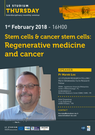
Prof. Marek Los

Speciality
Regenerative medicine cancer research
From
Jagiellonian University (Małopolska Center of Biotechnology) - PL
In residence at
Center for Molecular Biophysics (CBM) / CNRS - FR
Host scientist
Dr Catherine Grillon
PROJECT
Effects of electro-conductive, biomaterial-based tissue scaffolds on stem cells and transdifferentiation-derived somatic cells
Hypothesis: Combination of biodegradable, conductive polymers, and/or carbon-based nanomaterials, and/or bioactive elements, electro-stimulated would cause cyclical surface changes within scaffolds, that would stimulate iPS-differentiation into other progenitors and support transdifferentiation. The project focuses on the development of novel engineered artificial electro-conductive extracellular matrix materials, and characterization of their interactions with stem cells, both under normal condition and upon electro-stimulation. The engineered extracellular matrices will be obtained from combination of biodegradable polymers like i.e. PLGA, PCL, PLA with: (I) conductive polymers i.e. PPy, PEDOT, PANI, in order to make the combined polymers electro-conductive; (II) carbon-based nanomaterials, like single-wall carbon nanotubes (SWCNT), and/or multi-wall carbon nanotubes (MWCNT); (III) combination of conductive polymers, carbon-based, and bioactive elements, in order to achieve controlled drug release. Based on the above combinations of biomaterials (I, II and III), using electrospin, and 3D-printing, new composite biomaterials will be provided by collab. partner (Dr Hudecki, Ins. Nonferrous Metals, Gliwice, Poland). The biopolymers will be tested at the Centre de Biophysique Moléculaire, (Orleans, France), for (i) biocompatibility, (ii) induction of differentiation, (iii) combined effects of electro- and biostimulation, (iv) other effects like i.e. (un)desired phenotype drifting. The results will have a broad significance for science and the society: (a) reveal events at the interface between (tissue) stem cells and electro-conductive bioscaffolds mimicking nervous system processes; new insights, into engineering of biomaterials, nanotechnology, and regenerative medicine; (b) help define future directions for the development of new-generation biomaterials; (c) fostering scientific and cultural collaboration between Malopolska and Centre Val de Loire.
Publications
Adipose tissue is a promising source of mesenchymal stem cells. Their potential to differentiate and regenerate other types of tissues may be affected by several factors. This may be due to in vitro cell-culture conditions, especially the supplementation with antibiotics. The aim of our study was to evaluate the effects of a penicillin-streptomycin mixture (PS), amphotericin B (AmB), a complex of AmB with copper (II) ions (AmB-Cu2+) and various combinations of these antibiotics on the proliferation and differentiation of adipose-derived stem cells in vitro. Normal human adipose-derived stem cells (ADSC, Lonza) were routinely maintained in a Dulbecco’s Modified Eagle Medium (DMEM) that was either supplemented with selected antibiotics or without antibiotics. The ADSC that were used for the experiment were at the second passage. The effect of antibiotics on proliferation was analyzed using the 3-[4,5-dimethylthiazol-2-yl]-2,5-diphenyltetrazolium bromide (MTT) and sulforhodamine-B (SRB) tests. Differentiation was evaluated based on Alizarin Red staining, Oil Red O staining and determination of the expression of ADSC, osteoblast and adipocyte markers by real-time RT-qPCR. The obtained results indicate that the influence of antibiotics on adipose-derived stem cells depends on the duration of exposure and on the combination of applied compounds. We show that antibiotics alter the proliferation of cells and also promote natural osteogenesis, and adipogenesis, and that this effect is also noticeable in stimulated osteogenesis.
With the rapid advancement of regenerative medicine technologies, there is an urgent need for the development of new, cell-friendly techniques for obtaining nanofibers—the raw material for an artificial extracellular matrix production. We investigated the structure and properties of PCL10 nanofibers, PCL5/PCL10 core-shell type nanofibers, as well as PCL5/PCLAg nanofibres prepared by electrospinning. For the production of the fiber variants, a 5–10% solution of polycaprolactone (PCL) (Mw = 70,000–90,000), dissolved in a mixture of formic acid and acetic acid at a ratio of 70:30 m/m was used. In order to obtain fibers containing PCLAg 1% of silver nanoparticles was added. The electrospin was conducted using the above-described solutions at the electrostatic field. The subsequent bio-analysis shows that synthesis of core-shell nanofibers PCL5/PCL10, and the silver-doped variant nanofiber core shell PCL5/PCLAg, by using organic acids as solvents, is a robust technique. Furthermore, the incorporation of silver nanoparticles into PCL5/PCLAg makes such nanofibers toxic to model microbes without compromising its biocompatibility. Nanofibers obtained such way may then be used in regenerative medicine, for the preparation of extracellular scaffolds: (i) for controlled bone regeneration due to the long decay time of the PCL, (ii) as bioscaffolds for generation of other types of artificial tissues, (iii) and as carriers of nanocapsules for local drug delivery. Furthermore, the used solvents are significantly less toxic than the solvents for polycaprolactone currently commonly used in electrospin, like for example chloroform (CHCl3), methanol (CH3OH), dimethylformamide (C3H7NO) or tetrahydrofuran (C4H8O), hence the presented here electrospin technique may allow for the production of multilayer nanofibres more suitable for the use in medical field.
This study was designed to evaluate the relationship between Programmed cell death protein 6 (PDCD6) polymorphisms and cancer susceptibility. The online databases were searched for relevant case-control studies published up to November 2017. Review Manage (RevMan) 5.3 was used to conduct the statistical analysis. The pooled odds ratio (OR) with its 95% confidence interval (CI) was employed to calculate the strength of association. Overall, our results indicate that PDCD6 rs3756712 T>G polymorphism was significantly associated with decreased risk of cancer under codominant (OR = 0.82, 95%CI = 0.70–0.96, p = 0.01, TG vs TT; OR = 0.53, 95%CI = 0.39-0.72, p < 0.0001, GG vs TT), dominant (OR = 0.76, 95%CI = 0.66-0.89, p = 0.0004, TG+GG vs TT), recessive (OR = 0.57, 95%CI = 0.43-0.78, p = 0.0003, GG vs TT+TG), and allele (OR = 0.76, 95%CI = 0.67–0.86, p < 0.00001, G vs T) genetic model. The finding did not support an association between rs4957014 T>G polymorphism of PDCD6, and different cancers risk.
Lysosome‐associated protein transmembrane‐4 beta (LAPTM4B) has two alleles named as LAPTM4B*1 and LAPTM4B*2 (GenBank No. AY219176 and AY219177). Allele *1 has a single copy of a 19‐bp sequence in the 5` untranslated region (5`UTR), but allele *2 contains tandem repeats of 19‐bp sequence. LAPTM4B gene is located on long chromosome 8 (8q22.1) and contains seven exons that encodes two isoforms of tetratransmembrane proteins, LAPTM4B‐24 and LAPTM4B‐35, with molecular weights of 25 kDa and 35 kDa respectively. The LAPTM4B‐35′s primary structure is formed by 317 amino acid residues, and LAPTM4B‐24 comprised 226 amino acids. LAPTM4B, an integral membrane protein, contains several lysosomal‐targeting motifs at the C terminus and colocalizes with late endosomal and lysosomal markers. LAPTM4B is a proto‐oncogene, which becomes up‐regulated in various cancers. Preceding studies have examined the possible link between LAPTM4B polymorphism and susceptibility to several cancers,but the findings are still inconsistent. Hence, the present meta‐analysis was designed to investigate the impact of LAPTM4B polymorphism on risk of cancer.
The Rho GTPase family belongs to the Ras superfamily and includes approximately 20 members in humans. Rho GTPases are important in the regulation of diverse cellular functions, including cytoskeletal dynamics, cell motility, cell polarity, axonal guidance, vesicular trafficking, and cell cycle control. Changes in Rho GTPase signaling play an essential regulatory role in many pathological conditions, such as cancer, central nervous system diseases, and immune system-dependent diseases. The posttranslational modification of Rho GTPases (i.e., prenylation by mevalonate pathway intermediates) and GTP binding are key factors which affect the activation of this protein. In this paper, two essential and simple methods are provided to detect a broad range of Rho GTPase prenylation and GTP binding activities. Details of the technical procedures that have been used are explained step by step in this manuscript.
Stem cells are increasingly being used in the course of burn treatment. As several different types of stem cells are available for the purposes, it is important to chose the most efficient and the most practicable stem cell type. The aim of this study was to compare the potential of heterogeneous amnion cell mixture with the presently used standard therapy, the adipose tissue-derived stem cells. The placenta was collected during a Cesarean section procedure. Adipose tissue tissue-derived cells were isolated using the Cytori’s Celution® System. Cells were tested for fulfillment of the minimum criteria for stem cells. The efficiency of cell cultures was tested by an analysis of population doubling, cell proliferation, cell cycle and cell migration. Amniotic cells presented a higher ability for differentiation to chondrocytes and osteocytes than adipose-derived regenerative cells but a lower ability for differentiation toward adipocytes. Additionally, in vitro experiments have demonstrated a higher applicability of amniotic cells than adipose tissue-derived stem cells. Amniotic cells show several advantages: easy access to placenta, low costs and a lack of ethical dilemmas related to stem cell harvesting. The main disadvantage is, however, their availability, as isogenic treatment would only be possible for women around children-bearing age, unless personalized banks for amniotic cells would be established.
Final reports
ABSTRACT The combination of stem cell therapy with a supportive scaffold is a promising approach to improving tissue engineering. We aim producing novel material composites that may serve as artificial Extracellular Matrix (ECM). The natural ECM is composed of an organic (protein, polysaccharide) and inorganic (i.e. hydroxy-apatite) components that when combined with the cells form a tissue. ECM is an integral part of every tissue that besides providing the environment for cells to grow, it also improves tissue’s mechanical properties. It provides elasticity, flexibility and durability for the tissue. Tissue engineering approaches utilize artificial materials (biomaterials) as a substitute of natural ECM. The process of producing tissue scaffolds obtained from biodegradable polymers has become a very intensively researched area for the past several years. Most of the current work focuses on the design and preparation of scaffolds with use of various production technologies and different natural materials like chitosan, collagen, elastin and different synthetic ones, like polymer polycaprolactone (PCL), poly(lactic acid) (PLA), poly(ethylene oxide) (PEO). The objective of this study was to check the impact of the biomaterials on various cell types, and compare their growth pattern. Biodegradable PCL, and five of its hybrids: PCL+SHAP (SHAP, synthetic hydroxyapatite), PCL+NHAP (NHAP, natural hydroxyapatite), PCL+PLGA (PLGA, poly(lactide-co-glycolide), PCL+CaCO3, PCL+SHAP+NHAP+CaCO3 as well as one non degradable biomaterial: polyacrylonitryl (PAN), were tested. For the experiments four different cell types were used: human dermal skin fibroblasts, B16F10 (mouse melanoma cells), HSkMEC (Human Skin Microvascular Endothelial Cells) and HEPC-CB1 (Human Endothelial Progenitor Cells –Cord Blood 1). Impacts of the biomaterials on cells were assessed: 1) by measuring cytotoxic effect of the biomaterials liquid extracts and 2) by direct contact test. The ability of cells to attach to the biomaterials was tested as well as cells’ potential to growth and proliferate on the surface of the biomaterials. None of the tested biomaterials was cytotoxic towards the tested cells, making them a potential valuable raw ingredient for 3D scaffold development that would find its applications in tissue engineering. The differences in efficiency of cells attachment and proliferation between tested biomaterials and cells lines were observed. In addition, a stimulating effect of the biomaterials on cells growth was also detected.


